Back Of Neck Anatomy / Cervical Facet Joint Pain Treatment / See anatomy of the head and neck stock video clips.
Back Of Neck Anatomy / Cervical Facet Joint Pain Treatment / See anatomy of the head and neck stock video clips.. Headaches stemming from a neck problem are usually chronic and vary in type depending on the cause. In this video, i walk you through a basic approach to drawing the neck and upper back muscles. The content of the neck is grouped into 4 neck spaces, called the compartments. What bones do we have in our neck? The cervical spine supports the weight and movement of your head and protects the nerves exiting your brain.
The neck muscles, including the sternocleidomastoid and the trapezius, are responsible for the gross motor movement in the muscular system of the head and neck. The neck is the part of the body on many vertebrates that connects the head with the torso and provides the mobility and movements of the head. The neck is one of the most complex and intricate structures in our body and includes the spinal cord, which sends messages from the brain to the rest of the body. The neck is connected to the upper back through a series of seven vertebral segments. Muscle anatomy for bodybuilding 12 photos of the muscle anatomy for bodybuilding chest muscles anatomy for bodybuilders, muscle anatomy and bodybuilding, muscle anatomy for bodybuilding, muscle anatomy workout book, muscle anatomy workout pdf, human muscles, chest muscles anatomy for bodybuilders, muscle anatomy.
Posterior Cervical Anatomy | Free Images at Clker.com ... from www.clker.com The neck is connected to the upper back through a series of seven vertebral segments. The neck is essentially a passageway for air, food, liquids, blood, and more to travel between the head and the rest of the body, through structures such as blood vessels, nerves, and lymph nodes, as well as the larynx, trachea, and esophagus. The content of the neck is grouped into 4 neck spaces, called the compartments. This is a more stylized study and not meant to be entirely cor. Contains glands ( thyroid , parathyroid, and thymus ), the larynx , pharynx and trachea. Each nerve provides sensation to a specific area of the body called a dermatome. The cervical spine supports the weight and movement of your head and protects the nerves exiting your brain. The larynx is located where the pharynx, the back of the mouth and nasal cavity, divides into the trachea (the tube that carries air to the lungs) and the esophagus (the tube that carries food to.
In addition, there are also intervertebral joints.
The top of the cervical spine connects to the skull, and the bottom connects to the upper back at about shoulder level. In the neck are the thyroid and parathyroid glands, that secrete hormones that control metabolism and blood calcium levels. The neck is essentially a passageway for air, food, liquids, blood, and more to travel between the head and the rest of the body, through structures such as blood vessels, nerves, and lymph nodes, as well as the larynx, trachea, and esophagus. Think of it like a jigsaw puzzle, all the pieces fit in together and are required to get the full picture as to how it works. The back of the neck is mostly comprised of muscles, as well as the spine. They empty into the right and left subclavian veins in the base of the neck. The majority of these nerves control the functions of the upper extremities and allow you to feel your arms, shoulder, and back of your head. The neck muscles, including the sternocleidomastoid and the trapezius, are responsible for the gross motor movement in the muscular system of the head and neck. Vertebral, visceral and two vascular compartments. When talking about neck anatomy, we can't ignore the bones! The occipital bone is a bone that covers the back of your head; Headaches caused by a neck problem. The neurocranium (cranial vault) and the viscerocranium (facial skeleton).
The neurocranium (cranial vault) and the viscerocranium (facial skeleton). It consists of two major parts: Each nerve provides sensation to a specific area of the body called a dermatome. Muscle head anatomy vocal organ diagram female neck anatomy neck wireframe head neck human anatomy head artery anatomy face pharynx vector neck degree head anatomy 3d. The occipital bone is a bone that covers the back of your head;
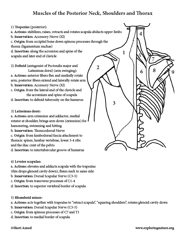
Muscles of the Neck, Shoulders, Chest and Thorax ... from www.exploringnature.org This is a more stylized study and not meant to be entirely cor. In the neck are the thyroid and parathyroid glands, that secrete hormones that control metabolism and blood calcium levels. Two of the main ligaments in the back are the anterior longitudinal ligament and the posterior longitudinal ligament. Each nerve provides sensation to a specific area of the body called a dermatome. The neurocranium (cranial vault) and the viscerocranium (facial skeleton). They move the head in every direction, pulling the skull and jaw towards the shoulders, spine, and scapula. The occipital bone is a bone that covers the back of your head; Anatomy of the back 12 photos of the anatomy of the back anatomy lungs back view, anatomy of a red back spider, anatomy of the back and kidneys, anatomy of the back neck muscles, anatomy of the human body back muscles, human anatomy, anatomy lungs back view, anatomy of a red back spider, anatomy of …
The superficial lymph nodes of the head and neck receive lymph from the scalp, face and neck.
The larynx is located where the pharynx, the back of the mouth and nasal cavity, divides into the trachea (the tube that carries air to the lungs) and the esophagus (the tube that carries food to. The occipital bone surrounds a large opening known as the foramen magnum. They empty into the right and left subclavian veins in the base of the neck. The four parathyroid glands are situated upon the back surface of the thyroid gland. The external carotid artery supplies the areas of the head and neck external to the cranium. An area called the occiput. It is made up of bones, discs, muscles, ligaments, nerves and tendons. The cervical spine, your neck, is a complex structure making up the first region of the spinal column starting immediately below the skull and ending at the first thoracic vertebra. Think of it like a jigsaw puzzle, all the pieces fit in together and are required to get the full picture as to how it works. This is a more stylized study and not meant to be entirely cor. The neck muscles, including the sternocleidomastoid and the trapezius, are responsible for the gross motor movement in the muscular system of the head and neck. When talking about neck anatomy, we can't ignore the bones! The neck is connected to the upper back through a series of seven vertebral segments.
The majority of these nerves control the functions of the upper extremities and allow you to feel your arms, shoulder, and back of your head. Seven vertebrae make up the cervical part of the spine, that is, the neck. The neck is connected to the upper back through a series of seven vertebral segments. The neck has seven to 10 ligaments, all serving a purpose in the structure and motion of the neck. Muscle anatomy for bodybuilding 12 photos of the muscle anatomy for bodybuilding chest muscles anatomy for bodybuilders, muscle anatomy and bodybuilding, muscle anatomy for bodybuilding, muscle anatomy workout book, muscle anatomy workout pdf, human muscles, chest muscles anatomy for bodybuilders, muscle anatomy.
Anatomy of the Spine Blog | Newark, New Jersey from www.suborthonj.com The majority of these nerves control the functions of the upper extremities and allow you to feel your arms, shoulder, and back of your head. Each nerve provides sensation to a specific area of the body called a dermatome. In this video, i walk you through a basic approach to drawing the neck and upper back muscles. The skeleton of the neck is composed of cervical vertebrae, the hyoid bone, the clavicles, and the sternum. Headaches caused by a neck problem. The structures of the human neck are anatomically grouped into four compartments; The internal jugular veins form the major venous drainage of the head and neck and are deep veins that parallel the common carotid artery. Neck anatomy nerves picture there are 8 spinal nerves that originate from the cervical spine.
After arising from the common carotid artery, it travels up the neck, passing posteriorly to the mandibular neck and anteriorly to the lobule of the ear.
They ultimately drain into the deep lymph nodes. The neck is one of the most complex and intricate structures in our body and includes the spinal cord, which sends messages from the brain to the rest of the body. Contains cervical vertebrae and postural muscles. What bones do we have in our neck? The skull is a strong, bony capsule that rests on the neck and encloses the brain. See anatomy of the head and neck stock video clips. The larynx is located where the pharynx, the back of the mouth and nasal cavity, divides into the trachea (the tube that carries air to the lungs) and the esophagus (the tube that carries food to. Contains glands ( thyroid , parathyroid, and thymus ), the larynx , pharynx and trachea. The external carotid artery supplies the areas of the head and neck external to the cranium. In the neck are the thyroid and parathyroid glands, that secrete hormones that control metabolism and blood calcium levels. The muscles of the back muscles make up a large part of the anatomy (structure) of the back. The internal jugular vein commences at the jugular foramen, and is the direct continuation of the sigmoid sinus, which is a large vein draining blood from the vein. The neck is the part of the body on many vertebrates that connects the head with the torso and provides the mobility and movements of the head.
0 Response to "Back Of Neck Anatomy / Cervical Facet Joint Pain Treatment / See anatomy of the head and neck stock video clips."


0 Response to "Back Of Neck Anatomy / Cervical Facet Joint Pain Treatment / See anatomy of the head and neck stock video clips."
Post a Comment